Lung abscesses can be classified based on the duration and the likely etiology. Acute abscesses are less than 4-6 weeks old, whereas chronic abscesses are of longer duration. Primary abscesses are infectious in origin, caused by aspiration or pneumonia in the healthy host. Secondary abscesses are caused by a preexisting condition (eg, obstruction), spread from an extrapulmonary site, bronchiectasis, and/or an immunocompromised state. Lung abscesses can be further characterized by the responsible pathogen, such as Staphylococcus lung abscesses and anaerobic abscess or Aspergillus lung abscess.
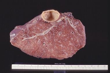
See the image below.

--> A thick-walled lung abscess.
In the 1920s, approximately one third of patients with lung abscess died. Dr David Smith postulated that aspiration of oral bacteria was the mechanism of infection. He observed that the bacteria found in the walls of the lung abscesses at autopsy resembled the bacteria noted in the gingival crevice. A typical lung abscess could be reproduced in animal models via an intratracheal inoculum containing, not 1, but 4 microbes, thought to be Fusobacterium nucleatum, Peptostreptococcus species, a fastidious gram-negative anaerobe, and, possibly, Prevotella melaninogenicus.
Lung abscess was a devastating disease in the preantibiotic era, when one third of the patients died, another one third recovered, and the remainder developed debilitating illnesses such as recurrent abscesses, chronic empyema, bronchiectasis, or other consequences of chronic pyogenic infections. In the early postantibiotic period, sulfonamides did not improve the outcome of patients with lung abscess. After penicillins and tetracyclines became available, outcomes improved. Although resectional surgery was often considered a treatment option in the past, the role of surgery has greatly diminished over time because most patients with uncomplicated lung abscess eventually respond to prolonged antibiotic therapy.
Most frequently, the lung abscess arises as a complication of aspiration pneumonia caused by mouth anaerobes. The patients who develop lung abscess are predisposed to aspiration and commonly have periodontal disease. A bacterial inoculum from the gingival crevice reaches the lower airways and infection is initiated because the bacteria are not cleared by the patient's host defense mechanism. This results in aspiration pneumonitis and progression to tissue necrosis 7-14 days later, resulting in formation of lung abscess.
Other mechanisms for lung abscess formation include bacteremia or tricuspid valve endocarditis causing septic emboli (usually multiple) to the lung. Lemierre syndrome, an acute oropharyngeal infection followed by septic thrombophlebitis of the internal jugular vein, is a rare cause of lung abscesses. The oral anaerobe F necrophorum is the most common pathogen.
Because of the difficulty obtaining material uncontaminated by nonpathogenic bacteria colonizing the upper airway, lung abscesses rarely have a microbiologic diagnosis.
Published reports since the beginning of the antibiotic area have established that anaerobic bacteria are the most significant pathogens in lung abscess. In a study by Bartlett et al in 1974, 46% of patients with lung abscesses had only anaerobes isolated from sputum cultures, while 43% of patients had a mixture of anaerobes and aerobes. [1] The most common anaerobes are Peptostreptococcus species, Bacteroides species, Fusobacterium species, and microaerophilic streptococci.
Aerobic bacteria that may infrequently cause lung abscess include Staphylococcus aureus, Streptococcus pyogenes, Streptococcus pneumoniae (rarely), Klebsiella pneumoniae, Haemophilus influenzae, Actinomyces species, Nocardia species, and gram-negative bacilli.
Two studies from Asia suggest that the bacteriologic characteristics of lung abscesses have changed. [2, 3] This finding is confirmed by a study performed by Takayanagi et al suggesting that Streptococcus species were the most common species, followed (in order of decreasing frequency) by anaerobes, Gemella species, and Klebsiella pneumoniae. These species were identified in this study with percutaneous ultrasonography-guided transthoracic needle aspiration and protected specimen brushes in a population of 205 patients.
Some geographic differences exist, with Streptococcus species being more prevalent in this study (done in one hospital in Japan), compared with previous accounts of anaerobic bacterial species being most predominant in Western populations. The study population notably had a 61% of individuals with periodontal disease, 16.6% were considered “alcoholic,” and 22.9% had significant diabetes mellitus. They were primarily Japanese, male (82%), smokers (75.6%), and alcoholic (34%). [3]
To support the findings by Takayangi et al, a subsequent study done by Want et al in a series of 90 patients with community-acquired lung abscess in Taiwan, anaerobes were recovered from just 28 patients (31%). The predominant bacterium was K pneumoniae, in 30 patients (33%). Another significant finding was that the rate of resistance of anaerobes and Streptococcus milleri to clindamycin and penicillin increased compared with previous reports. [2]
Both studies by Wang et al and Takayanagai suggest that aerobic organisms were more likely to be found in individuals with diabetes mellitus and periodontal disease, both risk factors for aerobic community acquired lung abscesses.
Nonbacterial and atypical bacterial pathogens may also cause lung abscesses, usually in the immunocompromised host. These microorganisms include parasites (eg, Paragonimus and Entamoeba species), fungi (eg, Aspergillus, Cryptococcus, Histoplasma, Blastomyces, and Coccidioides species), and Mycobacterium species.
The bacterial infection may reach the lungs in several ways. The most common is aspiration of oropharyngeal contents.
Patients at the highest risk for developing lung abscess have the following risk factors:
Periodontal disease Seizure disorder Alcohol abuseOther patients at high risk for developing lung abscess include individuals with an inability to protect their airways from massive aspiration because of a diminished gag or cough reflex, caused by a state of impaired consciousness (eg, from alcohol or other CNS depressants, general anesthesia, or encephalopathy).
Infrequently, the following infectious etiologies of pneumonia may progress to parenchymal necrosis and lung abscess formation:
Pseudomonas aeruginosa K pneumoniae S aureus (may result in multiple abscesses) Streptococcus pneumoniae Nocardia species Actinomyces species Fungal speciesPatients with COVID-19 who receive mechanical ventilation are at increased risk for development of lung abscessing pneumonia. [28, 30]
An abscess may develop as an infectious complication of a preexisting bulla or lung cyst.
An abscess may develop secondary to carcinoma of the bronchus. The bronchial obstruction causes postobstructive pneumonia, which may lead to abscess formation. Underlying lung cancer in edentulous patients with lung abscesses should also be considered.
There have been case reports of lung abscess associated with electronic cigarette use. [29, 32]
The frequency of lung abscesses in the general population is not known.
A male predominance for lung abscess is reported in published case series.
Lung abscesses likely occur more commonly in elderly patients because of the increased incidence of periodontal disease and the increased prevalence of dysphagia and aspiration. However, a case series from an urban center with high prevalence of alcoholism reported a mean age of 41 years. [4]
The prognosis for lung abscess following antibiotic treatment is generally favorable. Over 90% of lung abscesses are cured with medical management alone, unless caused by bronchial obstruction secondary to carcinoma.
Host factors associated with a poor prognosis include advanced age, debilitation, malnutrition, human immunodeficiency virus infection or other forms of immunosuppression, malignancy, and duration of symptoms greater than 8 weeks. [5] The mortality rate for patients with underlying immunocompromised status or bronchial obstruction who develop lung abscess may be as high as 75%. [6]
Aerobic organisms, frequently hospital acquired, are associated with poor outcomes. A retrospective study reported the overall mortality rate of lung abscesses caused by mixed gram-positive and gram-negative bacteria at approximately 20%. [7]
The mortality associated with an anaerobic lung abscess is lower than 15%, though it is slightly higher in patients with necrotizing anaerobic pneumonia and pneumonia caused by gram-negative bacteria. [32]
For patient education resources, visit the Infections Center and Lung Disease and Respiratory Health Center. Also, see the patient education articles Bacterial Pneumonia, Antibiotics, and Bronchoscopy.
A 54-year-old patient developed cough with foul-smelling sputum production. A chest radiograph shows lung abscess in the left lower lobe, superior segment.
A 42-year-old man developed fever and production of foul-smelling sputum. He had a history of heavy alcohol use, and poor dentition was obvious on physical examination. Chest radiograph shows lung abscess in the posterior segment of the right upper lobe.
A 42-year-old man developed fever and production of foul-smelling sputum. He had a history of heavy alcohol use, and poor dentition was obvious on physical examination. Lung abscess in the posterior segment of the right upper lobe was demonstrated on chest radiograph. CT scan shows a thick-walled cavity with surrounding consolidation.
Chest radiograph of a patient who had foul-smelling and bad-tasting sputum, an almost diagnostic feature of anaerobic lung abscess.